Phone: +7 (383) 330-67-71, Fax: +7 (383) 330-80-56, E-mail: bic@catalysis.ru
5 Lavrentiev Ave., 630090, Novosibirsk, Russia
Phone: +7 (383) 330-67-71, Fax: +7 (383) 330-80-56, E-mail: bic@catalysis.ru
5 Lavrentiev Ave., 630090, Novosibirsk, Russia
Morphology
Dispersed powder materials.
The following parameters are defined:
Jeol JEM-100CX and JEM-2010 electron microscopes (Japan) are designated for recording of transmission electron images with the magnification up to 1500 000 times. Accelerating voltage is 100 kV for JEM-100CX and 200 kV for JEM-2010. The lattice resolution is 0.2 nm at JEM-100CX, 0.14 nm at JEM-2010.
There are goniometers available for sample inclination up to 45° around the axes x and y and tomographic holder for the samples inclination up to 72° around the axe x. It used detachable device for the sample heating (up to 900°C) and cooling (to –170°C).
Electron microscopes are equipped with video-recording electron system, JEM-2010 is equipped by X-ray microanalyzer EDAX (EDAX Co) with semi-conductor detector, energy resolution is not worse than 130 eV. The elements from B to U are determined.
Dispersed sample in a quantity of several milligrams.
An example of reconstruction of the shape of metallic particle and carbon filament that formed on the particle in oxidation of hydrocarbons. The method of electron tomography (in High Angle Centered Dark Field regime).
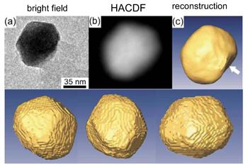 |
The model of the surface of metal particle (reconstruction) |
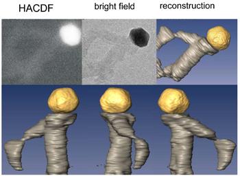 |
The model of carbon filament with metal particle (reconstruction) |
Dr. V.I. Zaikovskii. Catalysts and adsorbents.
Solid samples in the form of powder, plates, pellet and different things with size not more than 200 mm.
Jeol JSM-6460 LV scanning electron microscope that is modern digital model of the universal electron microscopy allows to get image of the solids surface with magnification by 5–300 000 times and resolution of 3 nm.
An original microscope design allows to use the full set of devices and options for solid surface study.
Results are presented in the following forms:
Sizes of solid sample are not more than 200 mm in diameter and 80 mm in height.
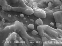 The surface of Ni3Al alloy |
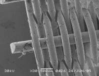 Netting of FeCrAl alloy |
Dr. A.N. Salanov. Solid samples.
The surface of solids with conductivity, including model metal supported catalysts, mainly – on carbon supports.
1 UHV scanning tunneling microscope (STM) GPI-300 combined with Auger spectrometer. The machine provides:
The best spatial resolution is: lateral – 0.03 nm, vertical – 0.002 nm (for specially prepared samples). The maximal scanning range is 1.2x1.2x1.0 μm3.
2 Ambient conditions scanning multi-microscope SMM2000 providing STM and Contact Mode Atomic Force Microscopy measurements.
The best spatial resolution is: lateral – 0.1 nm, in vertical direction – 0.02 nm (for specially prepared samples). The maximal scanning range is 30x30x2 μm3.
Electron conductivity of the sample surface for STM regime; the height variation across the scan frame – less than 2 μm.
Prof. V.I. Bukhtiyarov and Dr. R.I. Kvon. Model supported catalysts at carbon supports and planar oxide films.